Imaging Studies
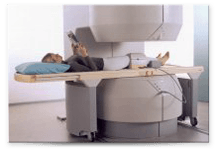
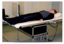
MRI PROCEDURE FOR HIPS
Patients are typically sent for specialized Magnetic Resonance Imaging (MRI) studies of the involved HIP (Not an MRI of the Pelvis).
Many times, patients are sent for an MRI of their painful hip but if the instructions are not very clear, they may wind up with the wrong study (i.e., an MRI of the pelvis, with special attention to the hip). Because this latter type of MRI is not sufficient, this common error results in overlooked problems and incorrect diagnoses. Additionally, many MRIs are performed on LOW field magnets (0.2 to 0.7 Tesla field strength magnets. These magnets are often done with body coils instead of SURFACE coils, and that results in a lower resolution, grainy and often inconclusive study.
Through our Hip Arthroscopy Center for Excellence, we aim to offer our patients the best MRI technology and care available. Our protocol includes at a minimum:
- MRI Arthrogram with local injection - 5cc of Marcaine.
- 1.5 – 3.0 Tesla Magnet preferred.
- MRI to be printed on films or CD
- Report is generated in 24 hours. Faxed directly to our office/emailed as well
- Films/CD given by the MRI facility to patient for MRI follow-up appointment
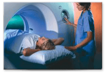
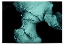
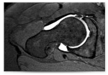
CT SCAN with 3-D RECONSTRUCTION
We follow a similar approach in standards of excellence for CT scan imaging. 3D CT scans are often necessary when assessing the shape of the femur (ball) and/or acetabulum (socket) in order to determine the degree of impingement and/or define the amount of bone that must be shaved to correct the underlying problem.
One of the most common reasons patients come to us after having had hip arthroscopy elsewhere is to correct underlying or unaddressed impingement, and the 3D CT scan helps define the best approach possible in this setting as well.
Through our Hip Arthroscopy Center for Excellence, we aim to offer our patients the best CT scan technology and care available. Our protocol includes at a minimum:
- CT Scans are always ordered with 3-D reconstruction
- Non-Contrast
- Printed on films or CD
- Report is generated in 24 hours. Faxed directly to our office/emailed as well
Films/CD given by the CT scan facility to patient for CT follow-up appointment
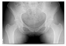
HIP X-RAYS
X-rays are requested along with the MRI/CT or in some cases they are repeated to compare to prior X-rays that may be outdated or incomplete. Our protocol includes:
- True AP Pelvis
- 'Frog Leg' Laterals both hips and in some cases Cross Table lateral X-rays as well
- Reports generated in 24 hours. Faxed directly to our office/emailed as well
- Films/CD given by the Radiology facility to patient for follow up appointment
When requesting a pre-visit consultation or second opinion, please make sure that you send the most recent X-rays, MRI, CT scan so that we may review your case with as much precision and accuracy as possible.